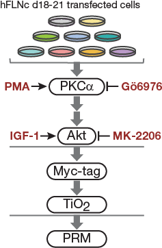
Warscheid Lab - Targeted FLNc S2233 S2236 phosphorylation assay

Targeted FLNc S2233 S2236 phosphorylation assay
  • Organism: Homo sapiens, Mus musculus
  • Instrument: Q Exactive Plus
  • SpikeIn: Yes
  • Keywords: FLNc, phosphoproteomics, PRM assay
  • Submitter: Lena Reimann
Abstract
In FLNc 10 phosphosites in FLNc (including S2234, S2237 and T2239) were found to be differentially phosphorylated. FLNc-pS2234 was by far the most abundant phosphosite followed by pS2237, while levels of FLNc-pT2239 were very low. pS2234 and pS2237 are part of the extended basophilic motif which is located in Ig-like domain 20 (d20)of FLNc. While levels of pS2234 and pS2234/pS2237 were strongly reduced following inhibition of PI3K/Akt signaling and increased upon IGF1-mediated stimulation, the levels were reversed for pS2237. This suggests that concurrent phosphorylation of FLNc at S2234 and S2237 prevails in vivo with pS2234 being the main site regulated by PI3K/Akt. In vitro kinase assays coupled to LC-MS (n=4) confirmed hFLNC-S2233 as prominent substrate site of Akt, while PKC alpha preferentially phosphorylateds S2236 in vitro. Accordingly, incubation with both kinases led to an increase of the doubly phosphorylated (pS2233 and pS2236) form. To validate these findings in myocytes, we performed an in cellulo kinase assays with pharmacologic interventions using Gö6976, PMA, IGF.1 and MK-2206, respectively, or a combination of the two inhibitors with PMA and IGF-1 followed by targeted MS analysis.
Experiment Description
Mouse C2 cells transiently expressing BirA* FLNc d18-21 were treated for 1 h with 10 µM MK-2206 (Selleck) or 10 µM Gö6976 (Merck) for 1 h or with 300 nM PMA (Sigma Aldrich) or IGF-1 (sigma Aldrich) for 15 min. In experiments using both, inhibitor and activator, cells were first treated with the respective inhibitor (MK-2206, Gö6976) for 1 h and PMA or IF-1 was added for the last 15 min of the treatment. Cells were lysed on ice with 500 µl modified RIPA buffer. Enrichment of the Myc-tagged FLNc fragment was performed with 10 µl Myc-Dynabeads (Invitrogen) according to the manufacturer’s protocol. On-bead tryptic digestion was performed in 200 µl ammonium bicarbonate solution (50 mM) for 3.5 h at 42°C and 1,400 rpm on a thermoshaker using sequencing grade trypsin,(Promega) in a 1:50 (w/w) ratio. Phosphopeptides were enriched as described above and mixed with 47.5-95 fmol phosphopeptides of a phosphopeptide standard comprising 181 phosphopeptides (Intavis). For targeted MS analysis on a QExactive Plus instrument, an inclusion list comprising 40 different precursor masses was generated with Skyline 2.6.0.6709. One scan cycle consisted of a full MS1 scan at a resolution of 70,000, an automatic gain control (AGC) target of 3e6 ions, a max. ion time of 20 ms and a scan range from m/z 400 to 1600, followed by 10 PRM scans. Each PRM scan targeted one precursor of the inclusion list at a resolving power of 17,500, an AGC target of 2e5, a max. ion time of 49 ms and an isolation window of 2 m/z. Raw files were analyzed with Skyline and intensities of hFLNc phosphopeptides were normalized to the summed intensities of eight phosphopeptides (SEGpSPVLPHEPAK, TGMGSGpSAGKEGGPFK, pSTVASMMHR, VIEDNEpYTAR, LIEDNEpYTAR, pSFNGSLKNVAVDELSR, pSGGQRHSPLSQR, AYpTHQVVTR) from the standard under control conditions.
Created on 3/4/20, 1:17 PM
Clustergrammer Heatmap
 
Download
FLNc_S2233_mutants_Phosphopeptides_2018-02-28_15-24-07.sky.zip2020-03-04 13:17:07912137812
PRMassay_FLNc+phosphostandard_2018-03-01_11-29-51.sky.zip2020-03-04 13:17:07913169624
PRMassay_FLNc+phosphostandard_pepmap.sky.zip2020-03-04 13:17:06912159022


Fig4C.

Cell-based kinase assay. PKC activator PMA, PKCa inhibitor Gö6976 and Akt activator IGF-1 and inhibitor MK-2206 were used to activate or block signaling pathways in C2 cells. For targeted MS analysis, phosphopeptides were enriched by Myc-tag and TiO2-based enrichment.

Figure 4D.

Immunoblot analysis of PKCα and Akt activities in C2 cells following pharmacologic interventions as indicated in 4C. Pan- and phospho-specific antibodies were used to detect total protein amounts and phosphoisoforms. GAPDH was used as loading control.
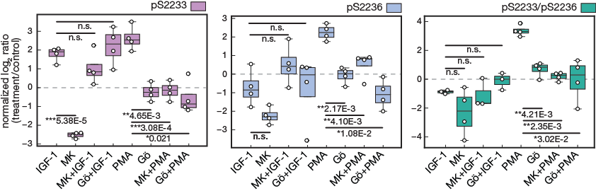
Figure 4e.

Targeted MS data of hFLNc phosphopeptides comprising the phosphorylation sites pS2233, pS2233/pS2236 and pS2236. MS data were quantified using Skyline and normalized to an internal phosphopeptide standard and the mock-treated control (DMSO). Intensities of phosphopeptides distinctive for a specific phosphorylation site in hFLNc d18-21 WT were added up per experiment and represented as normalized mean log2 ratio (treatment/control) ± SEM (n=4; *, p ≤ 0.05; **, p ≤ 0.01)


This data is available under the CC BY 4.0 license.